Scientific Illustrations
In my training as a biomedical communicator, I developed proficiency at communicating scientific content using a range of visual media.
Each project shown below involved a significant research stage to understand the content, and several design iterations before the final product was created.
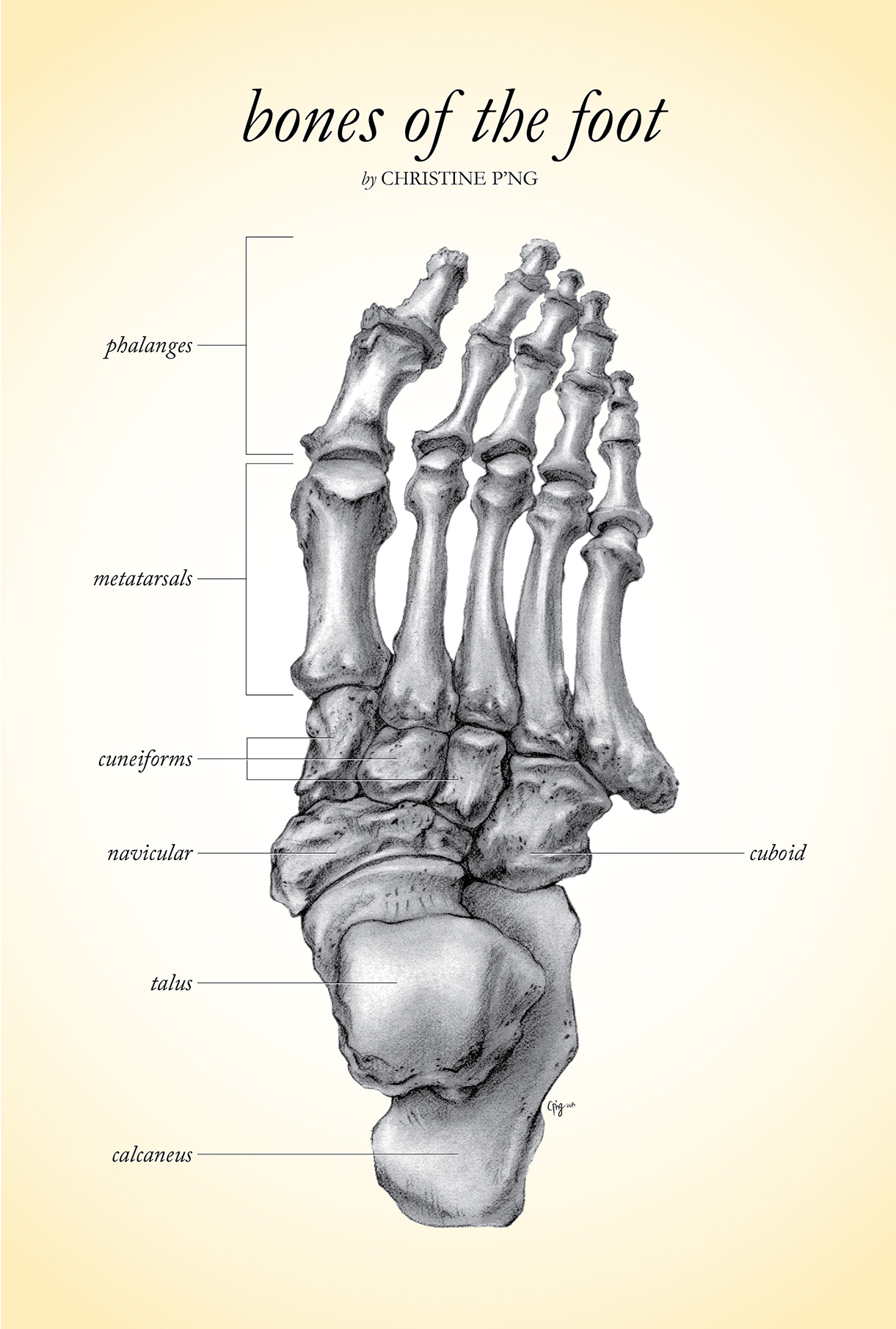
One of the first projects I created was a carbon dust drawing of the bones of the foot, created under the guidance of Professor Dave Mazierski at the University of Toronto.

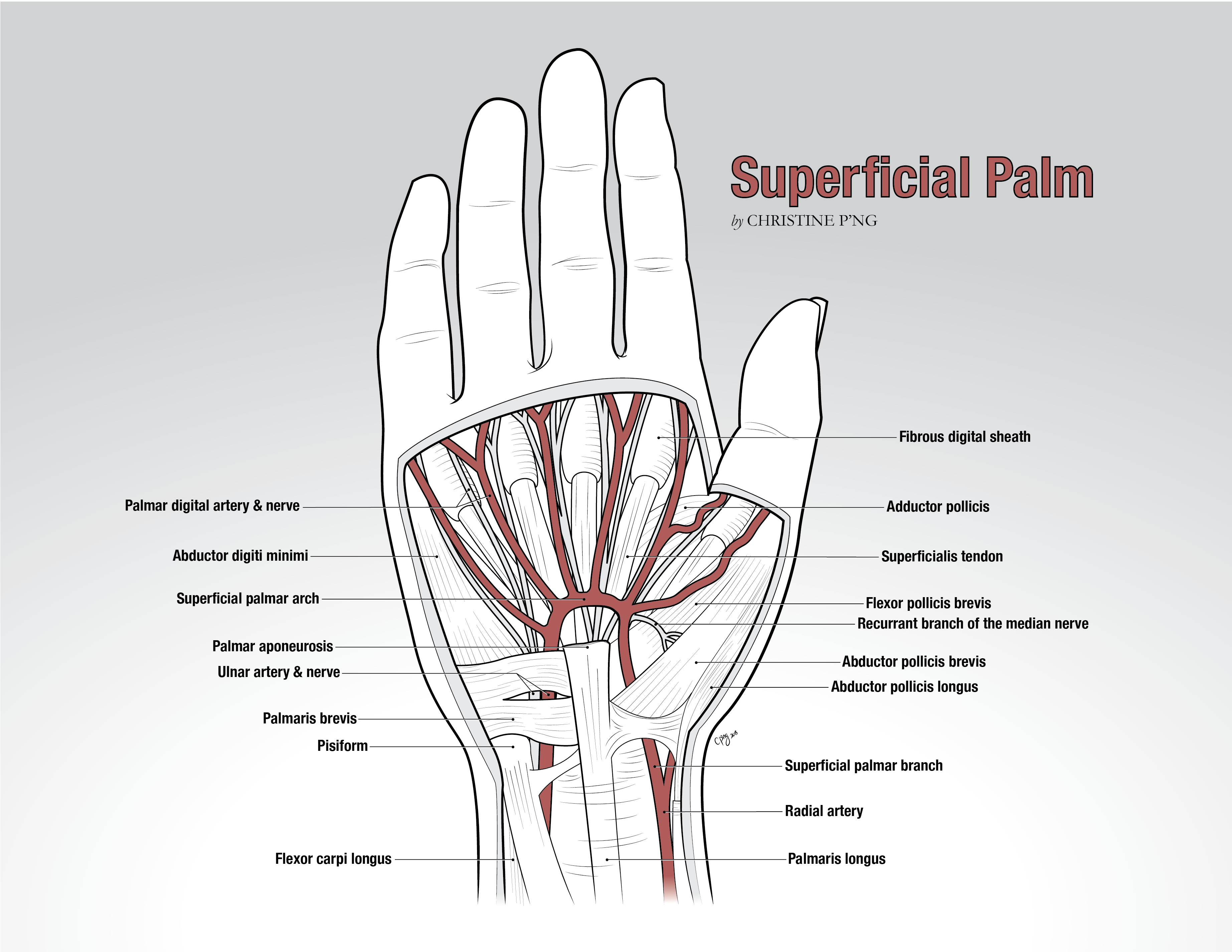
Line art is another common visual approach for conveying form, which I used to distinguish the many muscles, tendons, nerves, and blood vessels found in the superficial palm.

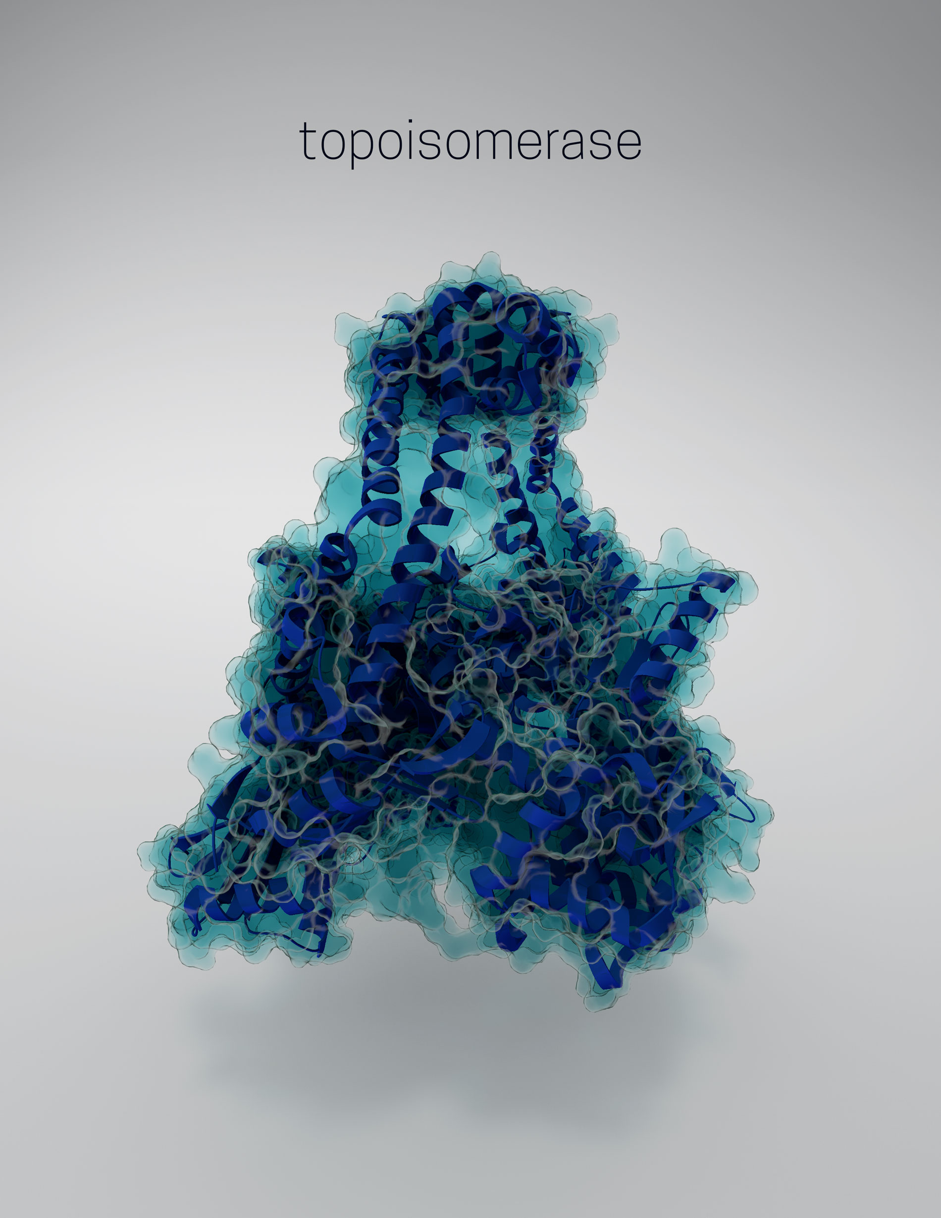
I also learned how to use 3D modelling software to illustrate scientific content. In this project I chose to model a human enzyme involved in DNA winding, topoisomerase. This project was created under the guidance of Professor Nick Woolridge, at the University of Toronto.
